| dc.contributor.author | Ocak, Süheyla | |
| dc.contributor.author | Yücel, Esra | |
| dc.contributor.author | Susam Şen, Hilal | |
| dc.date.accessioned | 2021-05-05T22:11:52Z | |
| dc.date.available | 2021-05-05T22:11:52Z | |
| dc.date.issued | 2020 | |
| dc.identifier.issn | 1300-0381 | |
| dc.identifier.uri | https://doi.org/10.5336/PEDIATR.2019-73027 | |
| dc.identifier.uri | https://hdl.handle.net/20.500.12933/203 | |
| dc.description | 2-s2.0-85092570018 | en_US |
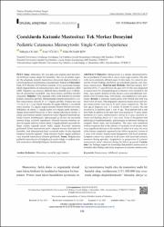
| dc.description.abstract | Objective: Mastocytosis is a disease characterized by the accumulation of mast cells in one or more organ systems. The skin is the most commonly affected organ. In this study, it is aimed to present the clinical findings and follow-up of pediatric patients with cutaneous mastocytosis. Material and Methods: Between January 2015 and June 2019, 27 cases between the ages of 0-18 who were diagnosed as mastocytosis by histopathological evaluation were included in the study. Age, gender, duration of rash, disease course and additional complaints were recorded, drug, food allergy, and anaphylaxis were questioned. Results: Cutaneous mastocytosis with limited skin rash was observed in all cases. Maculopapular cutaneous mastocytosis and solitary mastocytoma were seen in 24 and 3 cases, respectively. The median age at diagnosis was 11,5 months (3 months-12 years); the rash occurred in 26 patients under 1 year of age. Three patients had a rash from birth. In addition to the rash, there were pruritus in 11 cases, atopic dermatitis in 6 cases, asthma/reactive airway in 2 cases, recurrent urticaria and flashing attacks in 1 case each. None of the patients had pathological lymphadenopathy, splenomegaly or abnormal findings on complete blood count, and eosinophilia. The cases were monitored without treatment due to the absence of systemic involvement; there was a significant decrease in the number of rashes in 6 cases, but none of the lesions completely regressed in the follow-up period. Lesions of 3 cases with solitary mastocytoma disappeared with local treatment. Symptom control was achieved in all cases with local and systemic treatment during episodes. Anaphylaxis or progression of systemic mastocytosis wasn't observed during the follow-up period. Conclusion: Our findings support the knowledge that pediatric mastocytosis is limited to skin findings and rarely progresses to systemic mastocytosis. Copyright © 2020 by Türkiye Klinikleri. This is an open access article under the CC BY-NC-ND license (http://creativecommons.org/licenses/by-nc-nd/4.0/). | en_US |
| dc.language.iso | tur | en_US |
| dc.publisher | OrtadogŸu Reklam Tanitim Yayincilik Turizm Egitim Insaat Sanayi ve Ticaret A.S. | en_US |
| dc.rights | info:eu-repo/semantics/openAccess | en_US |
| dc.subject | Childhood | en_US |
| dc.subject | Cutaneous | en_US |
| dc.subject | Mastocytosis | en_US |
| dc.title | Pediatric cutaneous mastocytosis: Single-center experience [Çocuklarda Kutanöz Mastositoz: Tek Merkez Deneyimi] | en_US |
| dc.type | article | en_US |
| dc.department | AFSÜ, Tıp Fakültesi, Dahili Tıp Bilimleri Bölümü, Çocuk Sağlığı ve Hastalıkları Ana Bilim Dalı | |
| dc.contributor.institutionauthor | Susam Şen, Hilal | |
| dc.identifier.doi | 10.5336/PEDIATR.2019-73027 | |
| dc.identifier.volume | 29 | en_US |
| dc.identifier.issue | 3 | en_US |
| dc.identifier.startpage | 148 | en_US |
| dc.identifier.endpage | 152 | en_US |
| dc.relation.journal | Turkiye Klinikleri Pediatri | en_US |
| dc.relation.publicationcategory | Makale - Uluslararası Hakemli Dergi - Kurum Öğretim Elemanı | en_US |


