| dc.contributor.author | Çavuşoğlu, Dilek | |
| dc.contributor.author | Ataseven Kulalı, Melike | |
| dc.contributor.author | Olgaç Dündar, Nihal | |
| dc.contributor.author | Özer Gökaslan, Çiğdem | |
| dc.contributor.author | Aydın, Kürşad | |
| dc.date.accessioned | 2022-05-16T13:10:46Z | |
| dc.date.available | 2022-05-16T13:10:46Z | |
| dc.date.issued | 2022 | en_US |
| dc.identifier.citation | Çavuşoğlu, D., Ataseven Kulali, M., Olgaç Dündar, N., Özer Gökaslan, Ç., & Aydın, K. (2022). ITPA related developmental encephalopathy: Key role of neuroimaging. Annals of Indian Academy of Neurology. | en_US |
| dc.identifier.issn | 1998-3549 | |
| dc.identifier.uri | https://doi.org/10.4103/aian.aian_999_21 | |
| dc.identifier.uri | https://hdl.handle.net/20.500.12933/1019 | |
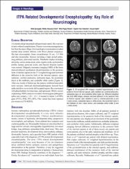
| dc.description.abstract | A 4-month-old girl presented with poor head control. She was born at term without complications. Parents were nonconsanguineous but from the same village. Her neurological examination revealed normal deep tendon reflexes with flexor plantar responses. She had microcephaly (head circumference 36 cm, -3.5 SD) and also dysmorphic features including a high arched palate, long philtrum, anteverted auricles. Metabolic studies including ammonia, serum amino acids, urine organic acids, acylcarnitine profile, lactate, pyruvate levels, and thyroid function studies were normal. Magnetic resonance imaging (MRI) of the brain showed delayed myelination (hyperintensities in the posterior limb of internal capsule) in the T2-weighted image and restricted diffusion in the posterior limb of the internal capsule, optic radiation, cerebral peduncles, substantia nigra, the pyramidal tracts in the midbrain, and cerebellar white matter [Figure 1]. After one month of follow-up, the patient exhibited intractable tonic seizures. Interictal electroencephalogram showed focal spike and slow waves in the left occipital region. She was treated with phenobarbital, levetiracetam, and topiramate. Whole–exome sequencing (WES) analysis revealed a homozygous pathogenic splice-site variant c. 124 + 1G > A located in intron 2 of ITPA gene (PVS1, PM2, PP3, PP5). This variant had been reported previously (rs376142053). | en_US |
| dc.language.iso | eng | en_US |
| dc.publisher | Medknow Publications | en_US |
| dc.relation.isversionof | 10.4103/aian.aian_999_21 | en_US |
| dc.rights | info:eu-repo/semantics/openAccess | en_US |
| dc.title | ITPA Related Developmental Encephalopathy: Key Role of Neuroimaging | en_US |
| dc.type | article | en_US |
| dc.authorid | 0000-0003-4924-5300 | en_US |
| dc.authorid | 0000-0001-6377-6162 | en_US |
| dc.authorid | 0000-0001-5345-1735 | en_US |
| dc.department | AFSÜ, Tıp Fakültesi, Dahili Tıp Bilimleri Bölümü, Çocuk Sağlığı ve Hastalıkları Ana Bilim Dalı | en_US |
| dc.contributor.institutionauthor | Çavuşoğlu, Dilek | |
| dc.contributor.institutionauthor | Ataseven Kulalı, Melike | |
| dc.contributor.institutionauthor | Özer Gökaslan, Çiğdem | |
| dc.identifier.volume | 25 | en_US |
| dc.identifier.issue | 1 | en_US |
| dc.identifier.startpage | 133 | en_US |
| dc.identifier.endpage | 134 | en_US |
| dc.relation.journal | Annals of Indian Academy of Neurology | en_US |
| dc.relation.publicationcategory | Makale - Uluslararası Hakemli Dergi - Kurum Öğretim Elemanı | en_US |


